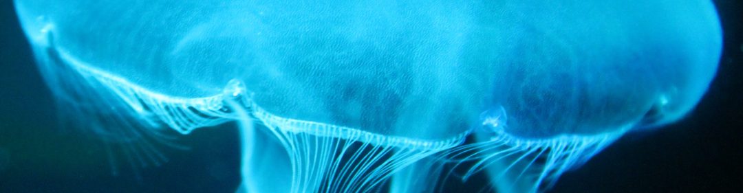
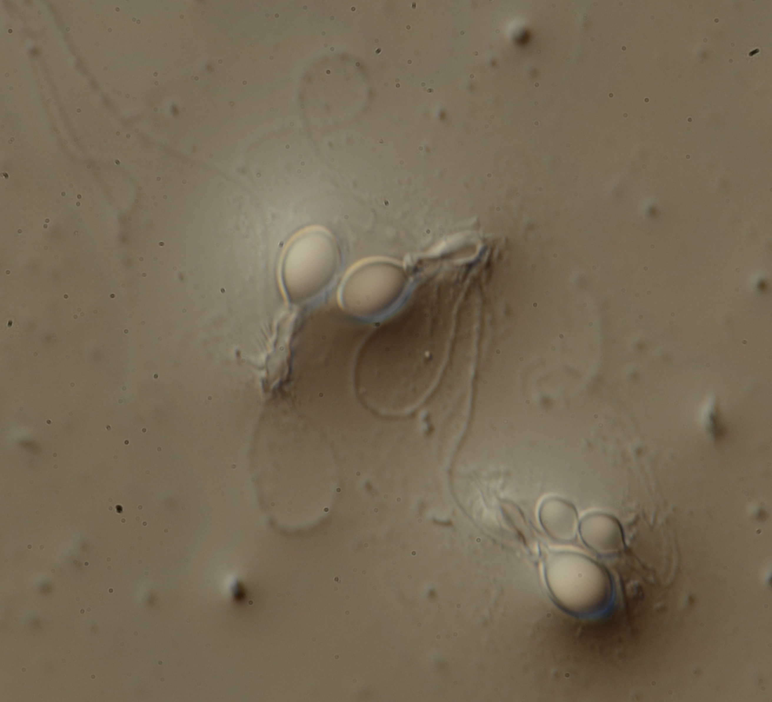
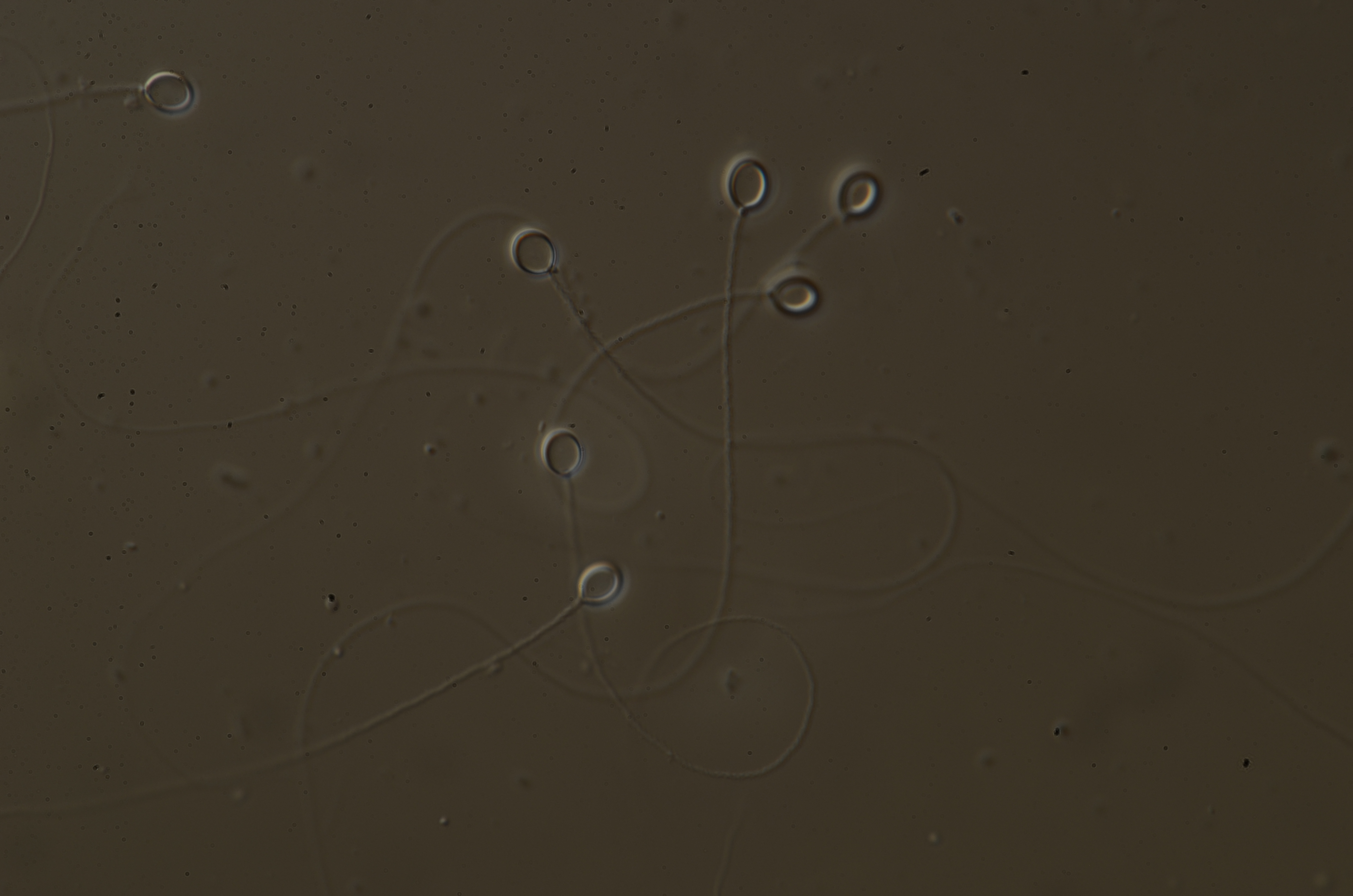
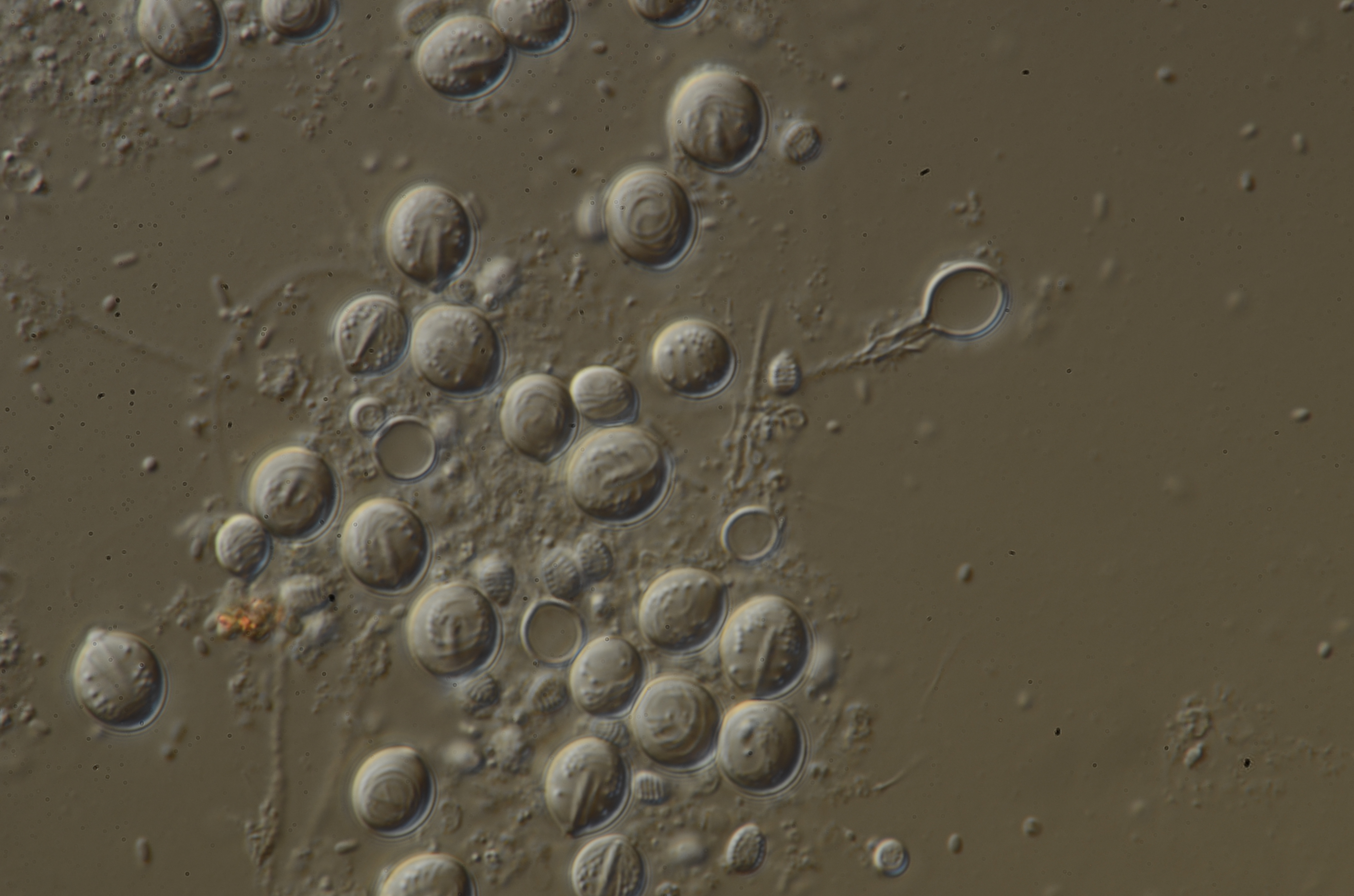
I am busy cranking away at the Smithsonian NMNH, but I wanted to share some photos of one of my favorite complex animal features: the stinging cell! Stinging cells house nematocysts, the stinging cell organelles distinctive to all cnidarians (hence the Latin translation, nettle-bearing animals). Nematocysts are thread-like capsules secreted by stinging cells that are mechanically or chemically discharged through a highly pressurized mechanism, releasing venom. Stinging cells come in over thirty different morphological types, but the nomenclature varies and new definitions are emerging all the time. But instead of trying to describe the extremely complex terminology, morphology, physiology, and evolutionary history of stinging cells right now (I could spend a whole doctorate thesis on any one of those), I wanted to show how freaking cool these structures are!


By the way, there is very little research on how these different morphological types correlate with venom composition. We know that specific types of nematocysts (stinging cell organelle) and nematocytes (stinging cells) are important taxonomically and functionally. That is, they can tell us about who is related to who evolutionarily and how specific kinds of stinging cell types are used for a specific purpose (prey capture, defense, competition, digestion, etc). But does morphology correlate with certain varieties of toxins deployed by these structures? Do highly penetrant cells with like birhopaloids and euryteles above contain more pore-forming toxins or proteases? Do the isorhizas contain more neurotoxins? Do stinging cells used for competition have a more potent sting than those used for general defense? Are there some stinging cells that contain little to no venom?
Even when I am counting and measuring a few thousand of these, it is not that hard to get caught up in how amazing these structures, and the animals that make them, really are.
All images taken by AK. Photo were taken in collaboration with Dr. Allen Collins (NOAA Systematics Lab) using animals from the Aquaroom located in the Invertebrate Zoology Division of the NMNH Smithsonian Institution. This project has been supported by a Lerner Gray Grant for Marine Science awarded by the ANMH and Graduate Studies funding from KU.
Definitions, for the bold:
Nematocyte: Stinging cell, contains nematocyst
Nematocyst: Stinging cell organelle, shared with all cnidarians, secreted from the Golgi apparatus of the nematocyte
Cnida (plural cnidae): Capsules (cells) that contain cnidairna intercellular secretory products, general term that includes cnidocysts, ptychocysts and spirocysts
Cnidoblast: Cells that makes the cnidae, general term that includes cnidoblasts, ptychoblasts, and spiroblasts
Cnidocyte: Mature cnidoblasts, general term that includes nematocytes, ptychocytes and spirocytes; interchangeable with nematocyst
Morphology:
Spines: Barbs along surface of the discharged nematocysts
Stylets: Large spines used to initially penetrate the target
Tubule: Thread-like structure that emerges from the capsule
Shaft: Enlarged portion at the end of the tubule near the capsule (stinging cell)
Haplonemes: Tubule lacking a shaft
Heteronemes: Tubule with well-defined shaft
Types mentioned above:
Birhopaloid: Two distinct dilations (bumps) along the shaft
Eurytele: Single dilation proximal along the shaft
Isorhiza: Roundish, uniform thickness of tubule; no dilations
References:
- Beckmann, A., & Özbek, S. (2012). The nematocyst: a molecular map of the cnidarian stinging organelle. International Journal of Developmental Biology, 56(6-7-8), 577-582.
- Corrales-Ugalde, M., Colin, S. P., & Sutherland, K. R. (2017). Nematocyst distribution corresponds to prey capture location in hydromedusae with different predation modes. Marine Ecology Progress Series, 568, 101-110.
- SmartEverDay. (2014). Jellyfish Stinging in MICROSCOPIC SLOW MOTION – Smarter Every Day 120. Retrieved from https://www.youtube.com/watch?v=7WJCnC5ebf4
- David, C. N., Özbek, S., Adamczyk, P., Meier, S., Pauly, B., Chapman, J., … & Holstein, T. W. (2008). Evolution of complex structures: minicollagens shape the cnidarian nematocyst. Trends in genetics, 24(9), 431-438.
- Fautin, D. G. (2009). Structural diversity, systematics, and evolution of cnidae. Toxicon, 54(8), 1054-1064.
- Heins, A., Glatzel, T., & Holst, S. (2015). Revised descriptions of the nematocysts and the asexual reproduction modes of the scyphozoan jellyfish Cassiopea andromeda (Forskål, 1775). Zoomorphology, 134(3), 351-366.
- Kass-Simon, G. S. A. A., & Scappaticci, Jr, A. A. (2002). The behavioral and developmental physiology of nematocysts. Canadian Journal of Zoology, 80(10), 1772-1794.
- Östman, C. (2000). A guideline to nematocyst nomenclature and classification, and some notes on the systematic value of nematocysts. Scientia Marina, 64(S1), 31-46.
